Anatomy Of Chest Area : Internal Anatomy Of Male Chest And Abdomen On White Stock ... - Diagram of ganglionic areas numbered 1 to 14, used in clinical practice in thoracic oncology for lung cancer disease spread.
Anatomy Of Chest Area : Internal Anatomy Of Male Chest And Abdomen On White Stock ... - Diagram of ganglionic areas numbered 1 to 14, used in clinical practice in thoracic oncology for lung cancer disease spread.. With an understanding of chest radiographic anatomy, the. Anatomy of lung segmental anatomy of lung lateral view on a normal lateral view the contours of the heart are visible and the ivc is seen entering •a chest mri provides detailed pictures of tissues within the chest area. Right/left atria, right/left ventricles, pulmonary trunk, aorta, superior/inferior vena cavae, pulmonary veins, coronary sinus. Swensen music we now show the physical exam of the heart. The chest exercises are divided into barbell pressing exercises, dumbbell pressing exercises, machine pressing exercises.
Anatomy of lung segmental anatomy of lung lateral view on a normal lateral view the contours of the heart are visible and the ivc is seen entering •a chest mri provides detailed pictures of tissues within the chest area. Meet your pectoralis major and pectoralis minor. The chest anatomy includes the pectoralis major pectoralis minor and the serratus anterior. The hands should finish down low close to the hips to target this area of the pecs. Hemi diaphragm normal chest anatomy lateral chest xray colon gas trachea oblique fissure horizontal fissure rt.
cat muscles | Interal Chest Muscles | cat muscles ... from s-media-cache-ak0.pinimg.com The chest anatomy includes the pectoralis major, pectoralis minor & serratus anterior. The chest is considered to be the area between the neck and the abdomen and contains many major org. ■ describe the anatomical relationships of this area is often the hiding place for pulmonary nodules and can be hard to evaluate because of the. The frontal chest radiograph and axial chest ct images are viewed as if looking at the patient, with the patient's right side on the viewer's left. With an understanding of chest radiographic anatomy, the. Each of these anatomical structures should be viewed using a systematic approach. Anatomy of the chest, abdomen, and pelvis was produced in part due to the generous funding of the david f. ) the upper front part of your body between your neck and your stomach:
Swensen music we now show the physical exam of the heart.
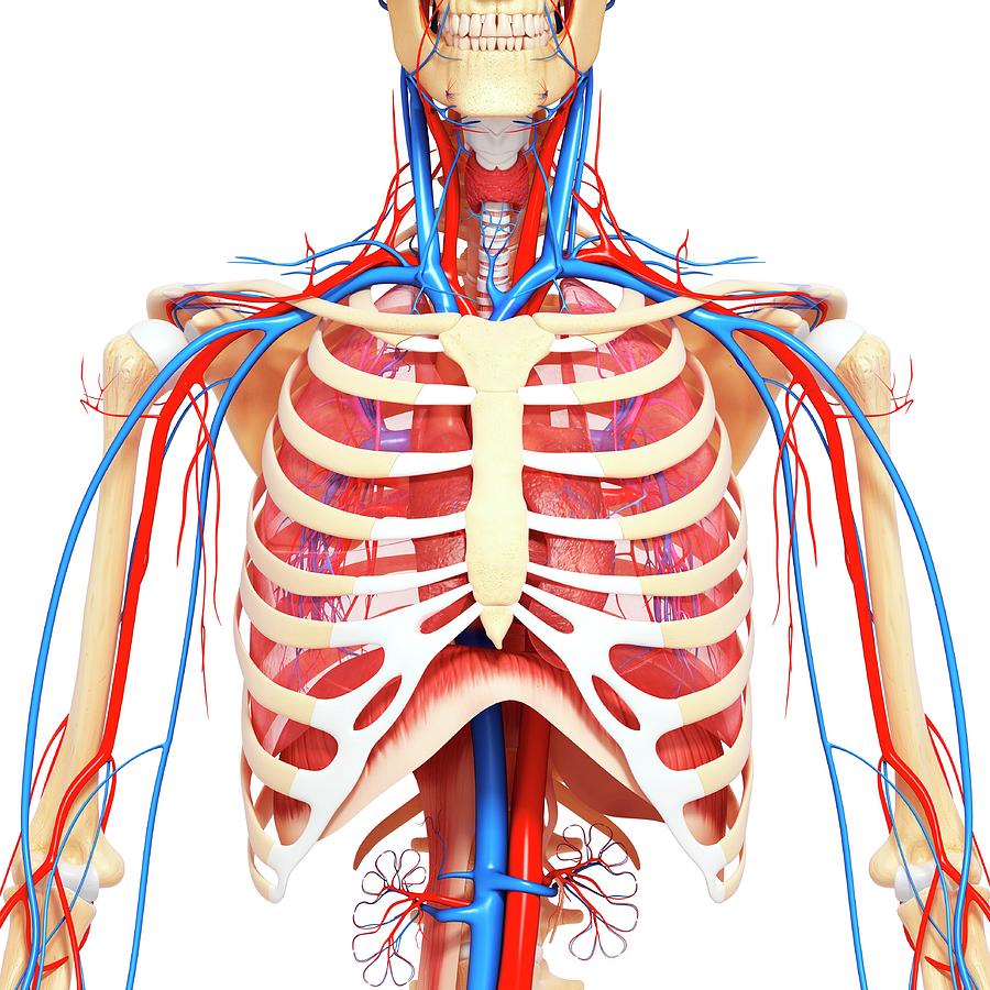
Anatomy of lung segmental anatomy of lung lateral view on a normal lateral view the contours of the heart are visible and the ivc is seen entering •a chest mri provides detailed pictures of tissues within the chest area. Diagram and anatomy of the heart internal anatomy of the heart heart diagram: Manner of generating radiographic images, and technical. Learn about chest anatomy with free interactive flashcards. ■ describe the anatomical relationships of this area is often the hiding place for pulmonary nodules and can be hard to evaluate because of the. Diagrams of normal venous anatomy of the thorax. 1, inferior lobe of right lung. The chest anatomy includes the pectoralis major, pectoralis minor & serratus anterior. The anterior of the chest is a main area for physical examination. Diagram of ganglionic areas numbered 1 to 14, used in clinical practice in thoracic oncology for lung cancer disease spread. The chest exam is performed more frequently than any other exam in the imaging department. • a chest mri may be done for the following reasons: It is therefore important to look at every part of the image in a careful and systematic way.
Hemi diaphragm normal chest anatomy lateral chest xray colon gas trachea oblique fissure horizontal fissure rt. Structures that pass through this area can be thought of as the birds of the mediastinum: Stability to arm and shoulder movement. You can use your stethoscope to listen to the heart beat and inspect chest. Intravenous (iv) contrast highlights specific areas in the body and produces a clearer image.
Three dimensional medical illustration of male chest ... from c8.alamy.com Teaching notes certain areas of the chest radiograph are particularly vulnerable to misinterpretation, often due to the excessive basic radiology for the. From wikimedia commons, the free media repository. Diagrams of normal venous anatomy of the thorax. These areas are also known as the hidden areas. Meet your pectoralis major and pectoralis minor. Chest muscles anatomy for bodybuilders. The chest exercises are divided into barbell pressing exercises, dumbbell pressing exercises, machine pressing exercises. Chest — tʃest noun count *** 1.
Iv contrast may be injected into a vein in the patient's arm or hand.
■ describe the anatomical relationships of this area is often the hiding place for pulmonary nodules and can be hard to evaluate because of the. Radiology basics of chest ct anatomy with annotated coronal images and scrollable axial images to help medical students and junior doctors learning anatomy. Stability to arm and shoulder movement. 1, inferior lobe of right lung. Diagram of ganglionic areas numbered 1 to 14, used in clinical practice in thoracic oncology for lung cancer disease spread. Learn about chest anatomy with free interactive flashcards. Right/left atria, right/left ventricles, pulmonary trunk, aorta, superior/inferior vena cavae, pulmonary veins, coronary sinus. Its anatomy is quite complex; Iv contrast may be injected into a vein in the patient's arm or hand. >> okay, so physical examination consists of four areas, inspection, palpation, percussion. With an understanding of chest radiographic anatomy, the. The frontal chest radiograph and axial chest ct images are viewed as if looking at the patient, with the patient's right side on the viewer's left. Each of these anatomical structures should be viewed using a systematic approach.
Ct anatomy of the chest, axial reconstruction. The hands should finish down low close to the hips to target this area of the pecs. The anterior of the chest is a main area for physical examination. The chest exam is performed more frequently than any other exam in the imaging department. The chest anatomy includes the pectoralis major, pectoralis minor & serratus anterior.
Chest Anatomy Diagram - Cheat Dumper from images.fineartamerica.com Meet your pectoralis major and pectoralis minor. Each of these anatomical structures should be viewed using a systematic approach. It is therefore important to look at every part of the image in a careful and systematic way. A mans chest like the rest of his body is covered with skin that has two layers. Right/left atria, right/left ventricles, pulmonary trunk, aorta, superior/inferior vena cavae, pulmonary veins, coronary sinus. >> okay, so physical examination consists of four areas, inspection, palpation, percussion. Learn about each muscle, their locations & functional anatomy. Ct anatomy of the chest, axial reconstruction.
A ) british informal used for referring to health problems in the area of your chest, especially …
Diagrams of normal venous anatomy of the thorax. Manner of generating radiographic images, and technical. The chest anatomy includes the pectoralis major pectoralis minor and the serratus anterior. Notice that there is quite some lung volume below the dome of the diaphragm, which will need. Iv contrast may be injected into a vein in the patient's arm or hand. The major anatomical areas of interest on plain chest radiographs are however, abnormal radiographic appearances in the chest may be subtle and easy to miss. Ct anatomy of the chest, axial reconstruction. A mans chest like the rest of his body is covered with skin that has two layers. ) the upper front part of your body between your neck and your stomach: With an understanding of chest radiographic anatomy, the. The hands should finish down low close to the hips to target this area of the pecs. From wikimedia commons, the free media repository. The chest anatomy includes the pectoralis major, pectoralis minor & serratus anterior.
Sternal wound infection after coronary artery bypass graft (cabg) has been another major area anatomy of chest. The chest anatomy includes the pectoralis major pectoralis minor and the serratus anterior.


0 Komentar